- Feb 18, 2025
Should You Trim or Extract? The Best Treatment for Rabbit Incisor Malocclusion
- Kristen Turner, DVM, DABVP (Exotic Companion Mammal Practice)
- 0 comments
Dental disease is one of the most commonly diagnosed conditions in exotic companion animal practice. One of the most prevalent findings is incisor malocclusion in rabbits. Keep reading for how I handle this frequent presentation in clinical practice.
Understanding Incisor Malocclusion in Rabbits
Incisor malocclusion has two underlying causes:
If the affected rabbit is less than 1 yr of age, there is a good chance this is congenital. Mandibular prognathism can result in fairly normal cheek teeth and maloccluded incisors at a young age
If the rabbit is older, this condition is almost certainly secondary to acquired dental disease (and it’s not uncommon for acquired dental disease to occur in young rabbits - so always evaluate the cheek teeth even if the patient is <1 yr of age).
Why Correct Diagnosis Matters for Treatment
Determining the underlying cause of the malocclusion is important for diagnostic, treatment and prognostic purposes. If the only problem is incisor malocclusion, extraction of all 6 incisors can be curative. If there is underlying cheek teeth disease, it must be managed - and this is often a lifetime disease.
Check out my previous blog on performing a rabbit physical exam for tips on performing a thorough oral exam.
Short-Term Management: Coronal Reduction (Incisor Trimming)
In the short term, coronal reduction (or trimming) of the incisors can be performed with a dremel or low-speed dental handpiece with either a cutting disc or dental burs (I like 701L cutting burs). Use caution when trimming the teeth - soft tissue damage can occur, or if the teeth are trimmed too short, the nerve can be exposed, causing pain and opening up potential for infection.
While this is a rapid procedure, it should be performed under sedation to minimize complication risk. For calmer rabbits, oral gabapentin +/- trazodone may be sufficient, but I also commonly use injectable sedation like butorphanol + midazolam if oral sedatives are not adequate.
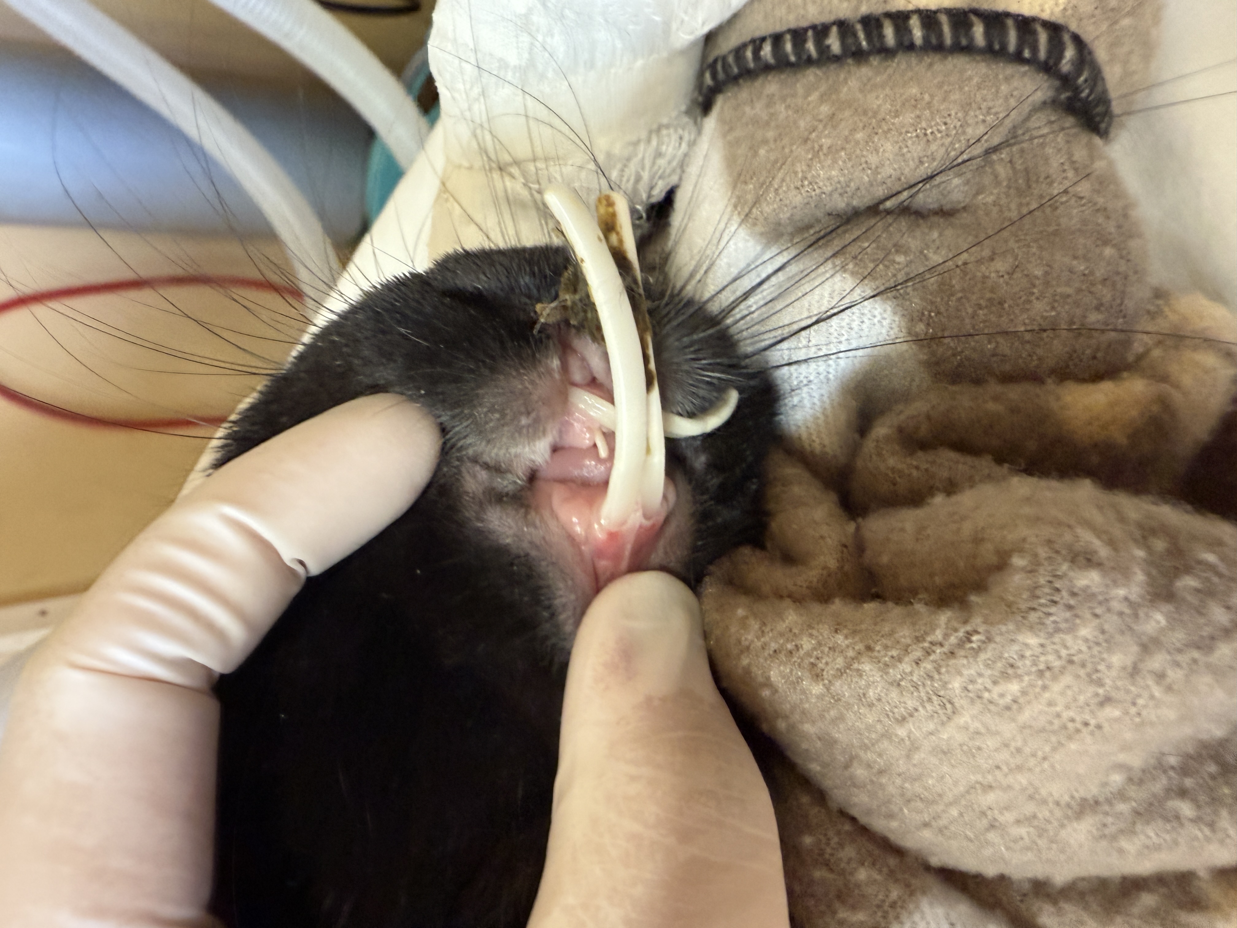
This is obviously not a rabbit :), but it demonstrates the concept. This sweet rat patient was sedated for coronal reduction with a low-speed handpiece and diamond disc, making sure to protect the soft tissues throughout the process.
The Importance of Extraction for Long-Term Treatment
Rabbit incisors grow extremely fast - up to 2 mm a week for the maxillary and 2.4 mm a week for mandibular incisors. This means that coronal reduction must be performed every 3-6 weeks depending on how severe the malocclusion is and if the overgrown teeth are impinging on soft tissues.
Because of this, extraction is always my treatment of choice!
This is a surgical procedure that most DVMs haven't learned in veterinary school, but it is very doable with a little bit of practice and familiarity with the anatomy! If you are able to practice the procedure during a wetlab or on a body donated to your clinic for scientific purposes, it greatly helps to understand the anatomy and the different forces that must be utilized to break down the periodontal ligaments.
Surgical Extraction of Rabbit Incisors: Step-by-Step Guide
-
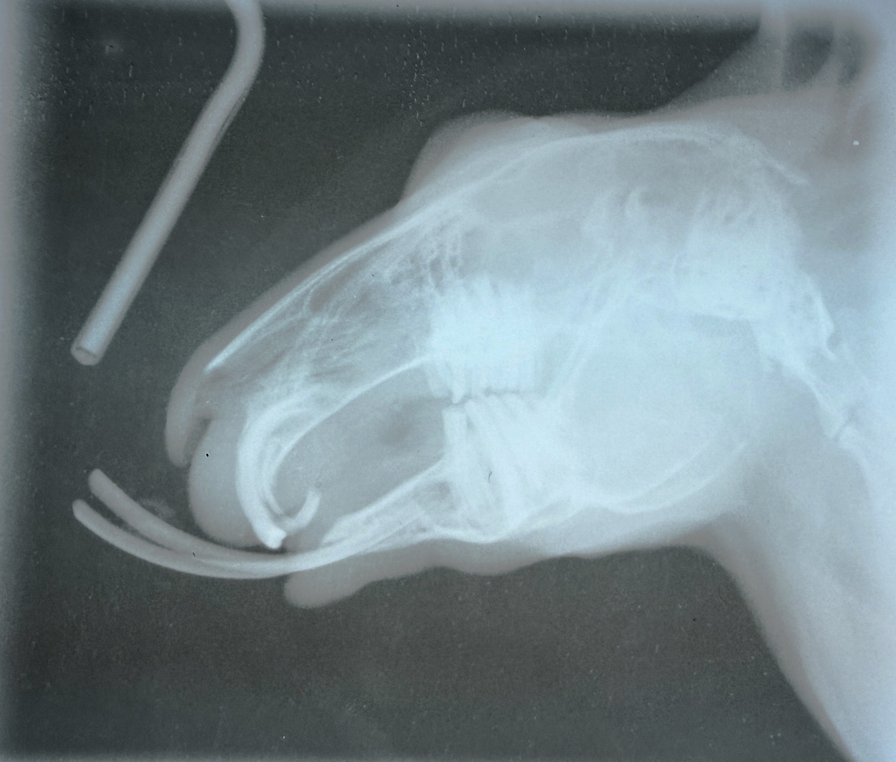
Once your patient is anesthetized, take skull radiographs to evaluate the apices of the teeth. In young rabbits, extraction is typically straightforward, however in older rabbits, the teeth can be abscessed or ankylosed to the surrounding bone. Radiographs help determine if extraction is possible. This is also a good opportunity to further evaluate the cheek teeth.
This is a film radiograph (what we can get in Belize!) and the technique isn't quite right - but it gives you an idea of how we can use radiographs to evaluate the curvature of the teeth, and check for the presence of sclerosis or apical lucency. It's a bit difficult to tell in this image, but there is a lucency at the base of the mandibular incisors, consistent with the abscess I found clinically on removal of the left mandibular incisor.
If a sedated oral exam and/or stomatoscopy has not been performed recently, use this opportunity to do a thorough oral exam.
Nerve blocks can be tricky in rabbits, but I attempt an infraorbital nerve block and an inferior alveolar nerve block with 1 mg/kg of lidocaine or bupivicaine. If you have access, I recommend potentiating your local anesthetic with either an opioid or dexmedetomidine for prolonged duration.
Gently flush the mouth and incisor teeth with an iodine-based antiseptic.
Use an 11 blade to incise around the attachments at the gingival margin of each tooth
-
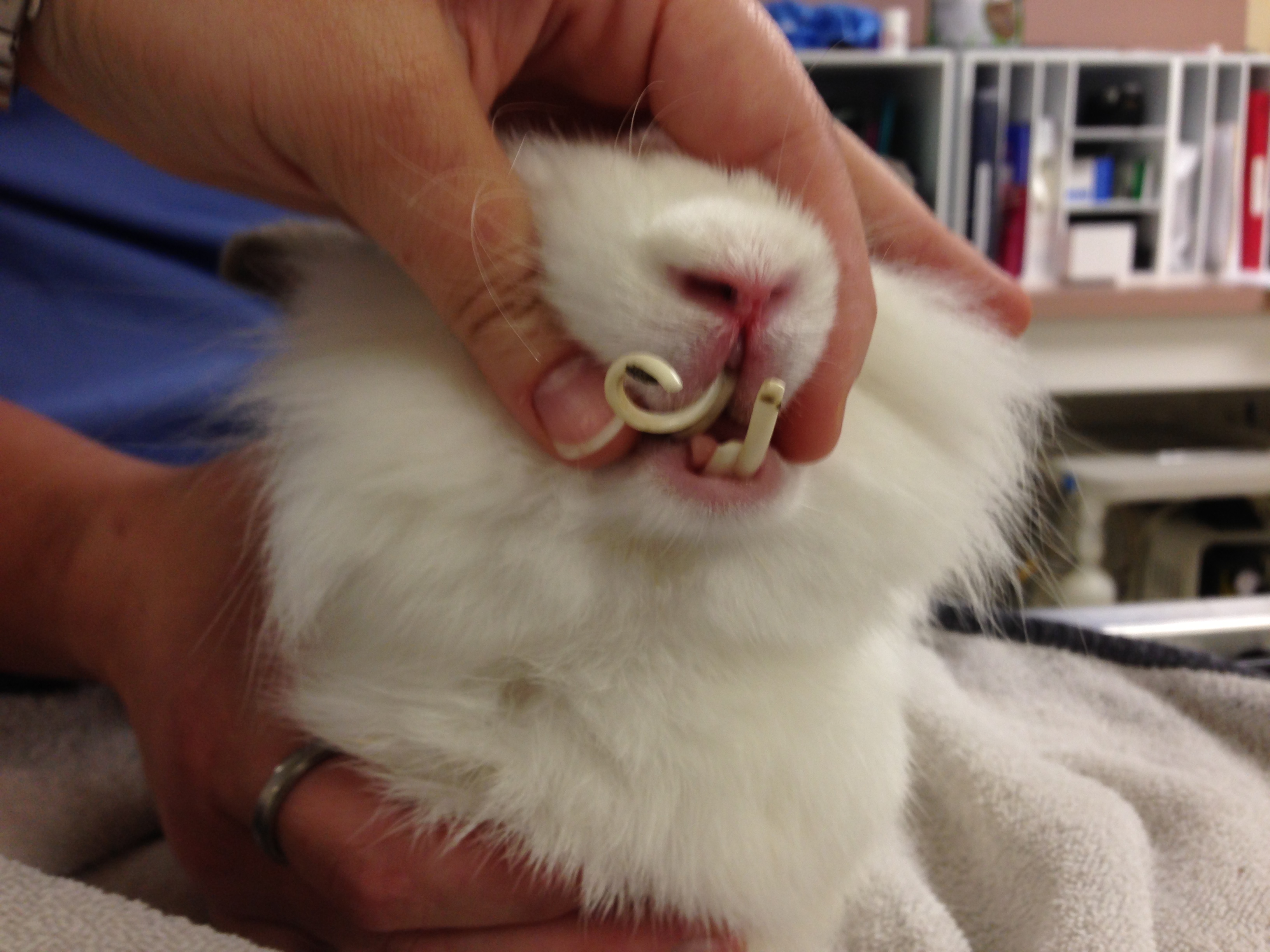
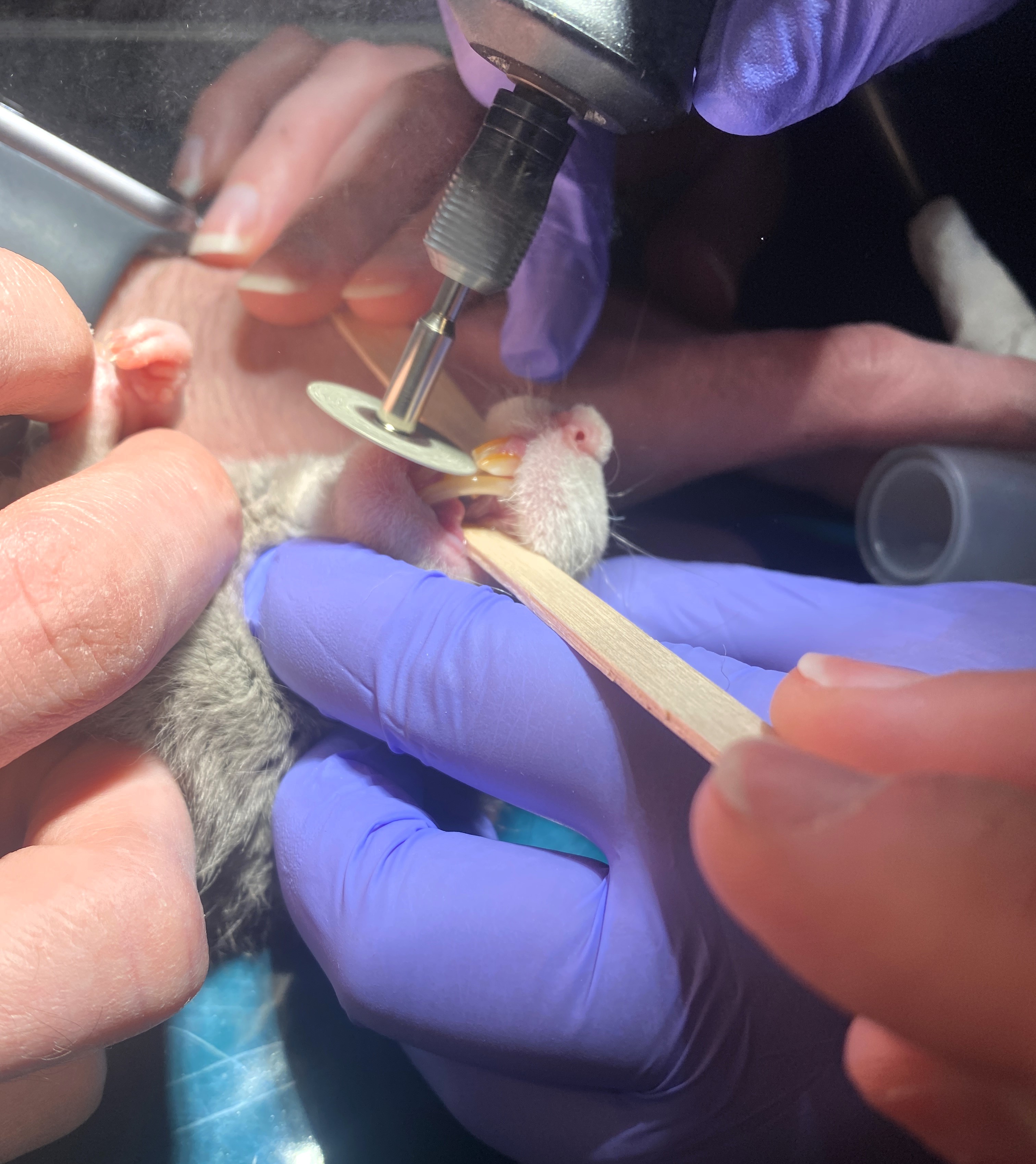
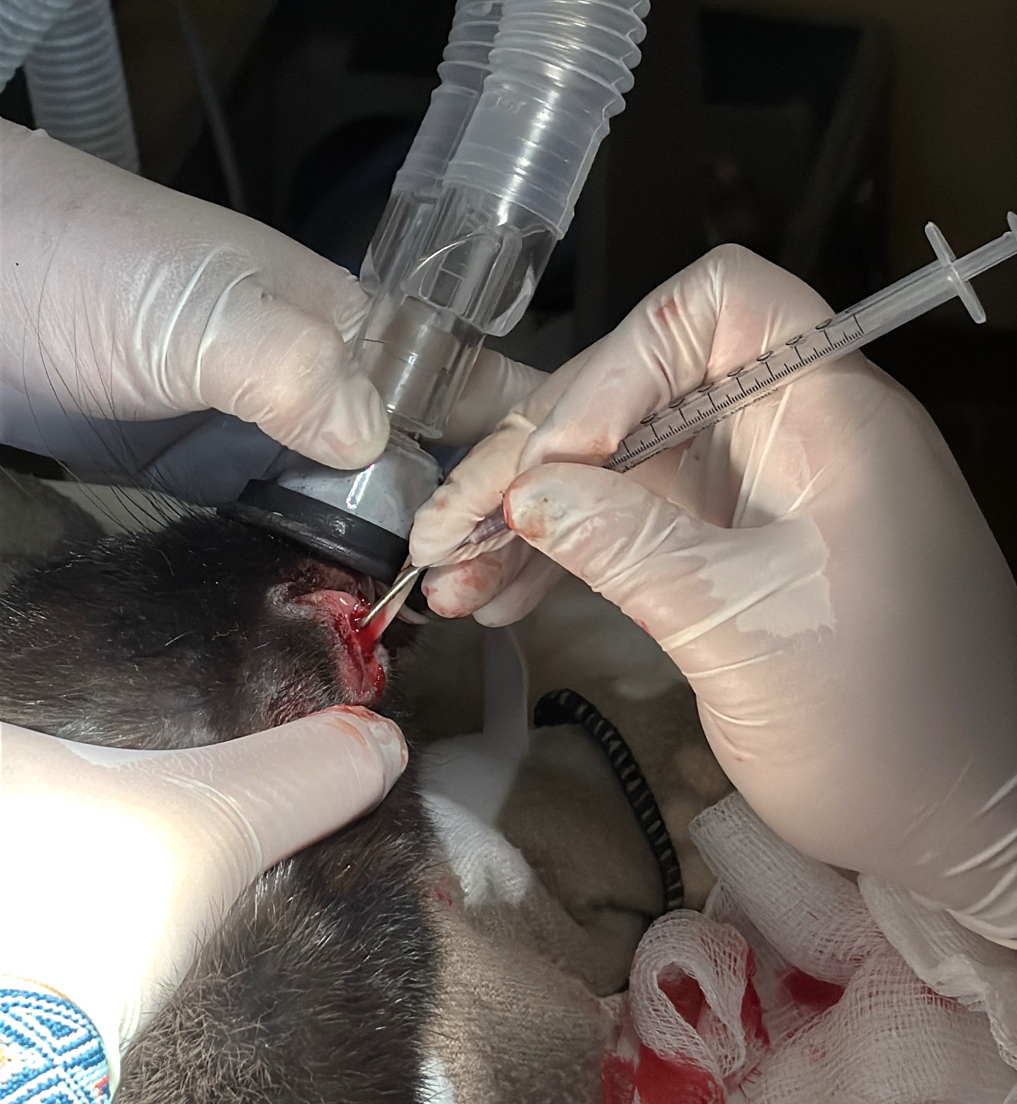
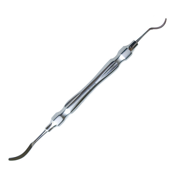
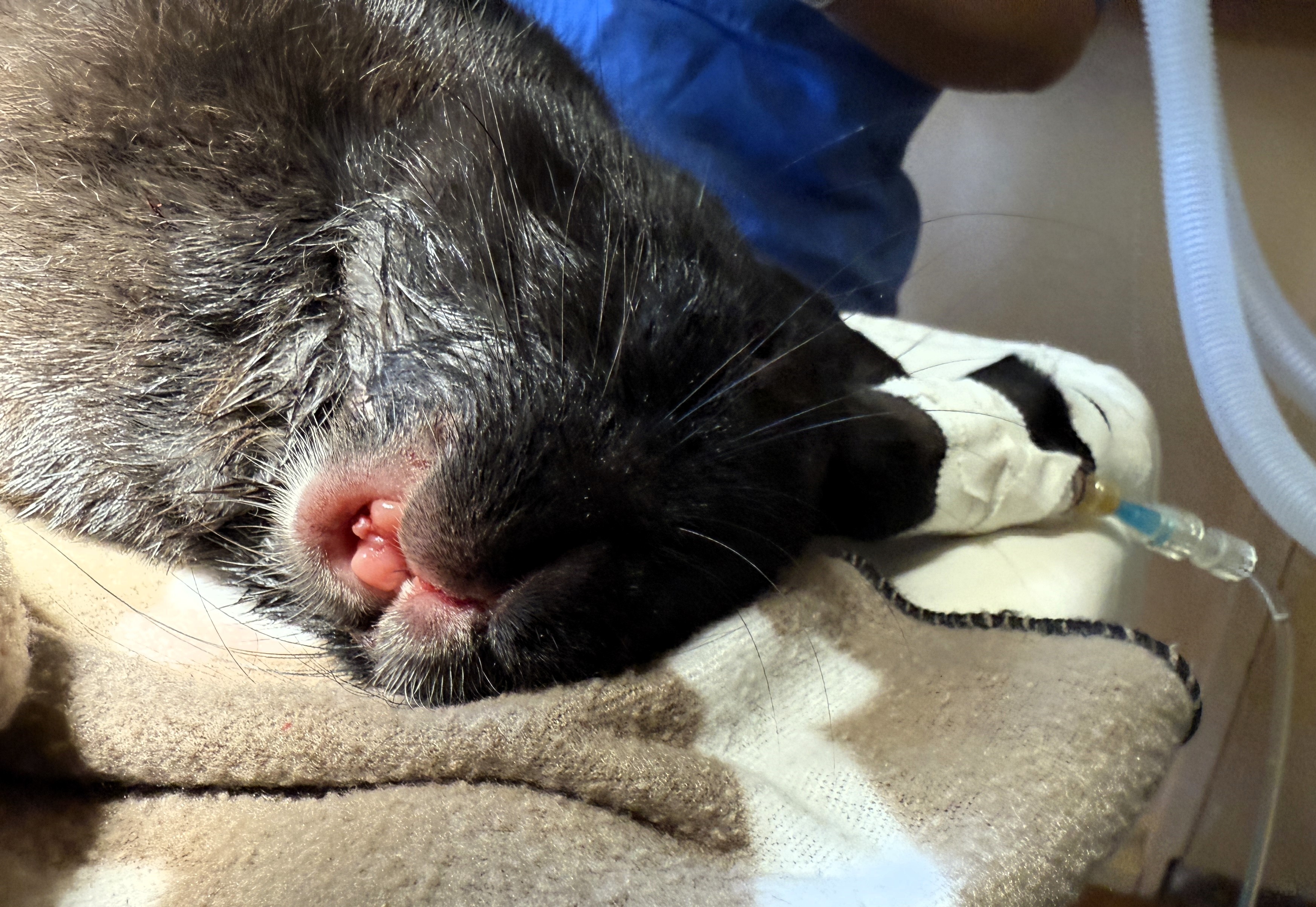
Use large hypodermic needles (18-20g) bent to match the curvature of the tooth to break down the periodontal ligament on each side of each tooth. Finer feline extractors can also be utilized as in the picture of the white rabbit above. A Crossley incisor elevator can then be utilized to further break the ligament down. Just like extractions in other species, this is a patience game. I work my way around each side of the tooth until it is nearly hanging out.
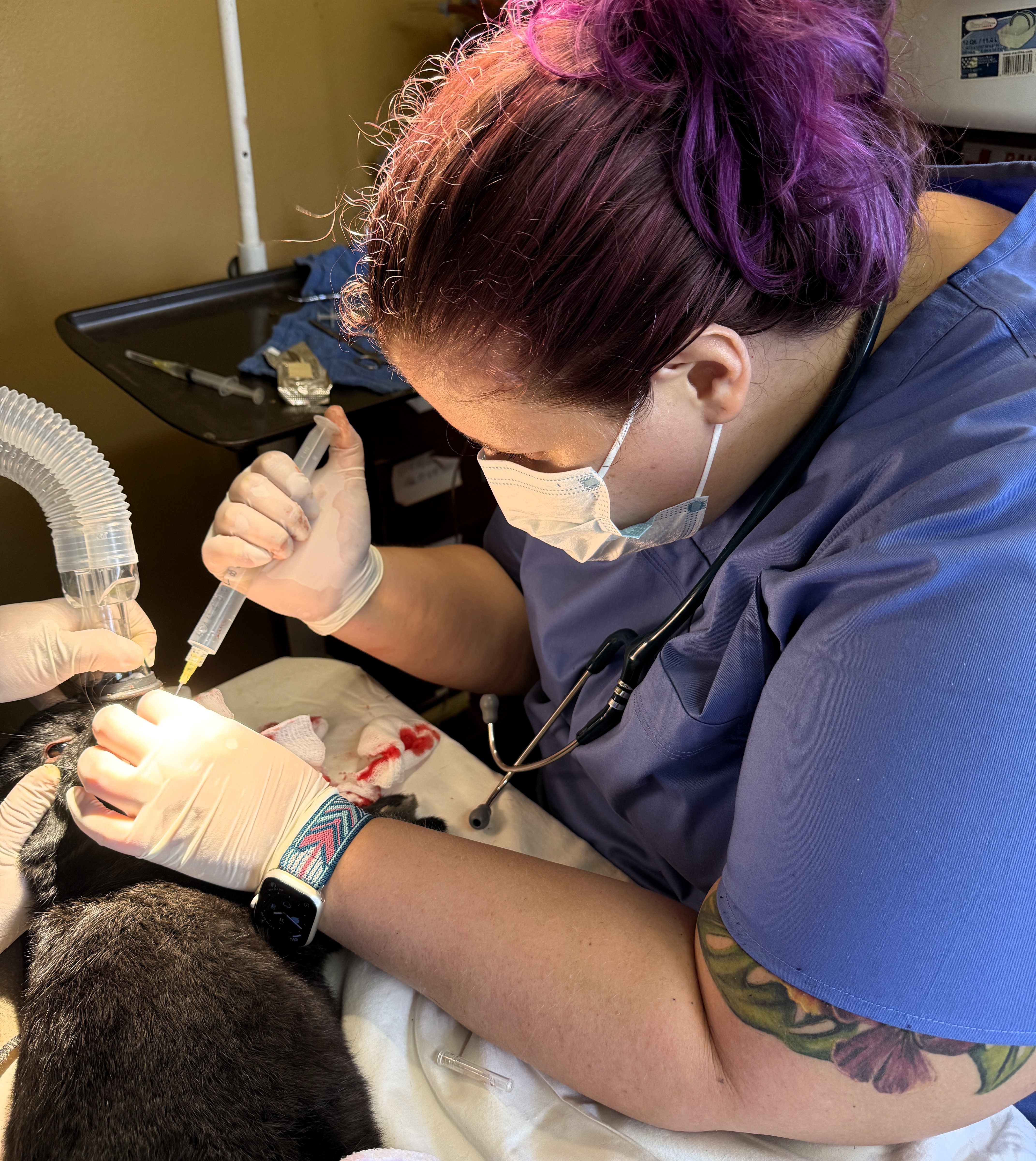
I always recommend intubating for dental procedures in rabbits, however I performed this procedure here in Belize and didn't have access to all the tools I typically utilize, like a rigid endoscope. In that case, we use what we have, and since rabbits are obligate nasal breathers, the nose can be masked for dental procedures (however, I strongly recommend intubation when possible as it is standard of care in most situations!).
Crossley Incisor Luxator from IM3 (multiple companies make a version!)
-
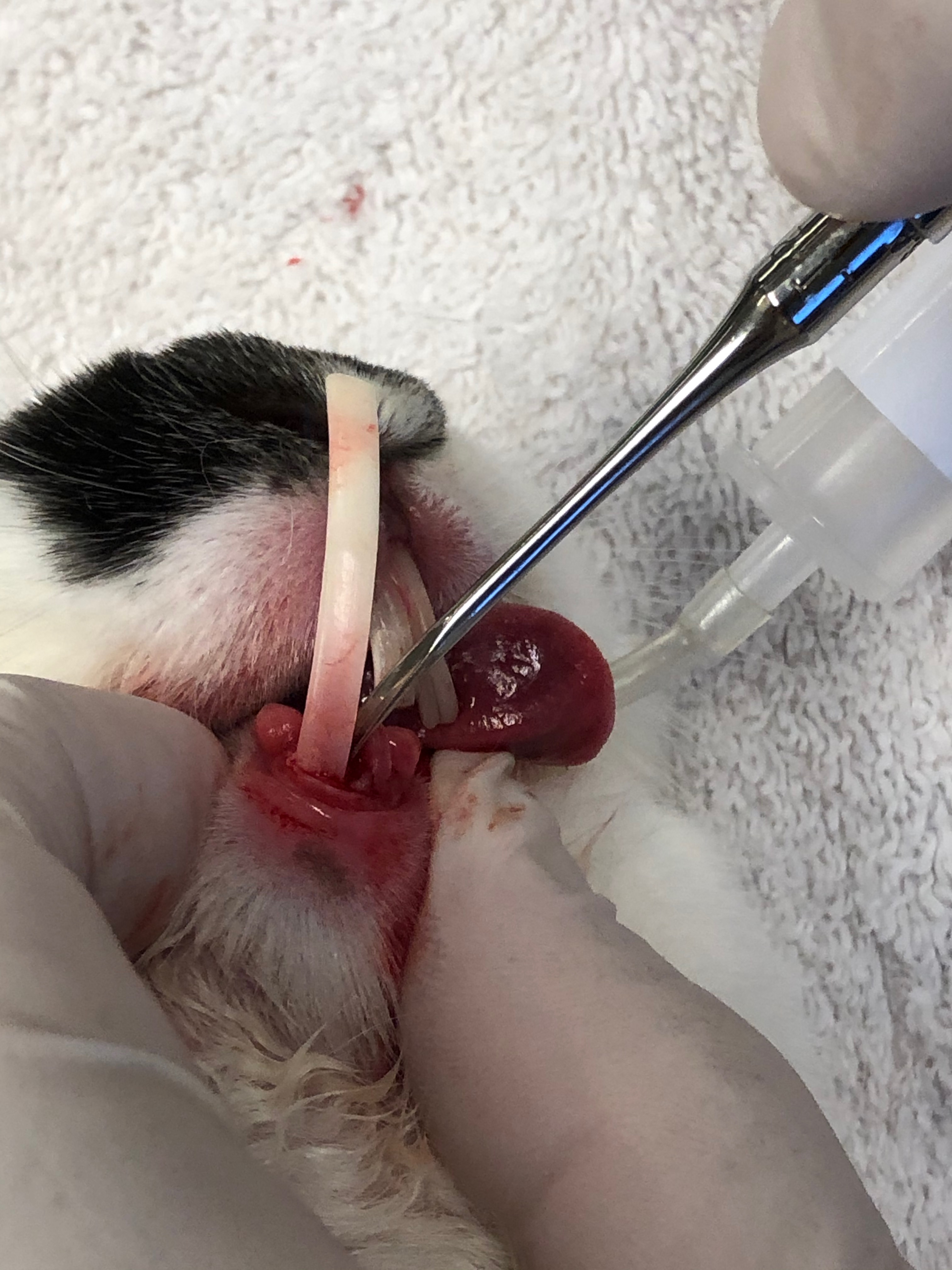
Once sufficiently loose, rock the tooth in the socket until it can be extracted. Before removing completely, push the tooth back into the socket and apply gentle pressure while rocking it back and forth for about 30 seconds - this helps destroy the germinal tissue. When you pull the tooth out, check the apex to make sure the tooth is complete and that pulp is present within the apex.
If there is evidence of apical infection, this is when I swab for culture & sensitivity or next-generation DNA sequencing.
You can also grab the apical tissue from the tooth that has been removed or a piece of the capsule if there is a defined abscess to send in with your sample.
I also use one of the bent hypodermic needles to traumatize the apical soft tissue to help prevent regrowth.
-
Once removed, flush the socket with sterile saline and/or dilute iodine solution.
Repeat for each tooth, including the smaller peg teeth.
-
Close the upper and lower gingiva with a purse-string suture around each opening. I like to use 4-0 or 5-0 absorbable monofilament (usually poliglecaprone 25) and try to get at least 4-6 points of contact.
-
If a tooth is abscessed, I leave that extraction site open to allow for continued flushing and drainage
In this patient I closed the upper gingiva but the left mandibular incisor was abscessed so I left the mandibular extraction sites open.
-
-
I recheck in 7-10 days to ensure the extraction sites are healing well. Sutures will dissolve and fall out on their own.
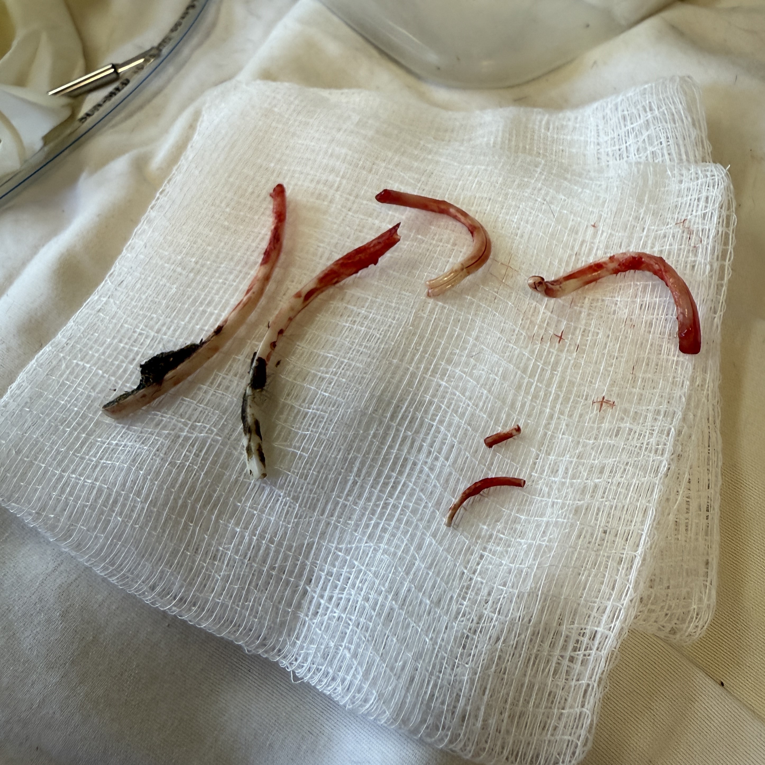
All 6 extracted teeth from the black rabbit shown above on a 4x4 gauze. Note the corkscrew-like curvature of the maxillary incisor. Also note that one peg tooth is smaller than the other - in this case, I believe the tooth was growing abnormally as it was adjacent to the corkscrew tooth and there appeared to be pulp present in the removed portion, however I advised the owner that it was possible the tooth had fractured and could regrow if there was any germinal tissue left behind.
Post-Extraction Care and Healing
I ensure that we are providing the rabbit with adequate analgesia throughout the procedure, utilizing an opioid pre-med, local block and potentially a continuous rate infusion of additional analgesics (lidocaine +/- ketamine +/- opioid). This helps prevent wind-up pain and most of my patients are eating almost immediately once they are alert.
I send home on meloxicam 1 mg/kg q12hrs for 3 days (if renal function is adequate) and then q24hrs until recheck. I will also add gabapentin or buprenorphine if I feel like the extractions were especially traumatic or painful.
I always send home with a small amount of a critical care herbivore diet and instruct the owners to supplement q8hrs if the patient is not eating. However, I’ve found most of the time they are eating by the time they go home.
Potential Complications and How to Address Them
It’s important to warn the owner that even if everything is done 100% correctly, regrowth can occur. If that happens, it’s typically only 1 tooth, and extraction can be attempted again in the future. Sometimes severely ankylosed pieces of tooth may be impossible to remove without further imaging like CT. Some teeth even need to be removed via an extraoral approach through the mandible or a rhinotomy.
Final Thoughts: Setting Rabbits Up for Long-Term Dental Health
Incisor malocclusion in rabbits is more than just an overgrown tooth problem - it’s often a sign of deeper dental disease. While coronal reduction (trimming) can provide temporary relief, extraction of all six incisors is the best long-term solution to help keep rabbits comfortable and avoid future potential complications like abscess formation. A proper diet rich in hay and low in pellets is also crucial for preventing future dental issues.
Got questions or experiences with rabbit incisor malocclusion? Drop them in the comments or send me a message - I’d love to hear from you!
Stay in the loop on exotic veterinary medicine
Join my email list to get weekly newsletters to keep you in-the-know in exotic vet med and be the first to receive access to new resources, courses, masterclasses, and more!